|
Tools for Biomedical Research
 At the forefront of biomedical research, medical scientists use particle accelerators to explore the structure of biological molecules. They use the energy that charged particles emit when accelerated to nearly the speed of light to create one of the brightest lights on earth, 30 times more powerful than the sun and focused on a pinpoint.
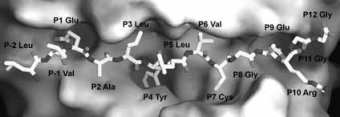
Deciphering the structure of proteins is key to understanding biological processes and healing disease. To determine a protein's structure, researchers direct the beam from an accelerator called a synchrotron through a protein crystal. The crystal scatters the beam onto a detector. From the pattern of scattering, computers calculate the position of every atom in the protein molecule and create a 3-D image of the molecule.
Physicists originally built synchrotron accelerators to explore the fundamental nature of matter. At first, they looked on synchrotron radiation as a troublesome problem that sapped electrons' acceleration energy. However, they soon saw the potential to use this "nuisance" energy to create ultrapowerful beams to study biological molecules and other materials.
Now, researchers at synchrotron light sources use dedicated particle accelerators to explore the molecules of life with matchless power and precision. Future accelerators will create still higher-energy beams for both particle physics and biomedicine.
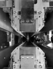 An undulator, in use at the Advanced Light Source at DOE's Lawrence Berkeley National Laboratory. Each undulator contains two 4.55-meter-long arrays of permanent magnets with alternating polarity.The arrays are supported by a superstructure capable of resisting the force of their attractionóup to 42 tons (the weight of a 38,000 kg mass). As an electron beam passes through a vacuum chamber between the arrays, the magnets cause the beam to curve back and forth and thus to produce synchrotron radiation. Undulators produce light brighter than that from other types of synchrotron radiation sources, with the added characteristics of partial coherence and linear polarization. In this photograph, a strobe light emulates the electron beam.
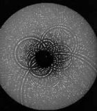 Diffraction image of a biomolecule. Beams from a synchrotron light source pass through crystals of protein to create images like this one. Using this diffraction technique, scientists can use light sources from particle accelerators to decode the internal structure of complex protein molecules such as enzymes.
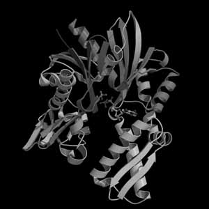
 Researchers using a beamline at the Advanced Photon Source at DOE's Argonne National Laboratory have discovered clues that promise a better understanding of the prevention of juvenile diabetes. Here, an insulin molecule binds to a human glycoprotein found at the cell surface.
|
