Seeing beneath the surface
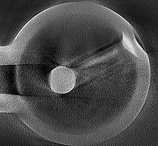 |
This is a 3.9-GHz higher-order mode coupler. Image courtesy of North Star Imaging |
|
|
X-ray tomography gives subsurface view of accelerator cavities
The power of the CAT scan to find tumours or bone fractures has advanced medical science by bringing such ailments to X-ray light, foregoing the need to cut a body open for diagnosis. As particle accelerator research makes its own advances, scientists are exploiting the X-ray in similar ways.
Researchers at the US Department of Energy Fermi National Accelerator Laboratory have been using X-ray computed tomography (CT) to take their search for faults in accelerator cavities and associated structures beneath the surface. Penetrating X-rays can reveal performance-limiting cracks and holes that would otherwise go undetected.
“It’s been a really cool tool for us,” said Fermilab’s Elvin Harms. Unlike its most familiar application in the medical field, X-ray tomography isn’t used to to view a cavity’s innards – there’s nothing in a cavity but empty space. But flaws and other weakness can hide within a cavity’s walls or other places, such as cavity end groups, that are tough to access without breaking cavities apart.
The method of diagnosis of a cavity by X-rays is fundamentally the same as that of a patient in a CAT (computed axial tomography) scanner. It begins by placing the suspected-broken structure inside a radiation-safe X-ray cabinet, between the X-ray tube and a digital detector. The differences in material thickness and density attenuate the X-rays differently.
Read more
—Leah Hesla
|